Blanching Lesion Meaning
When you press on it it stays red and does not lighten or turn white blanch. Since they happen in so many different places and types of tissue they have many different causes and means of diagnosis and treatment.
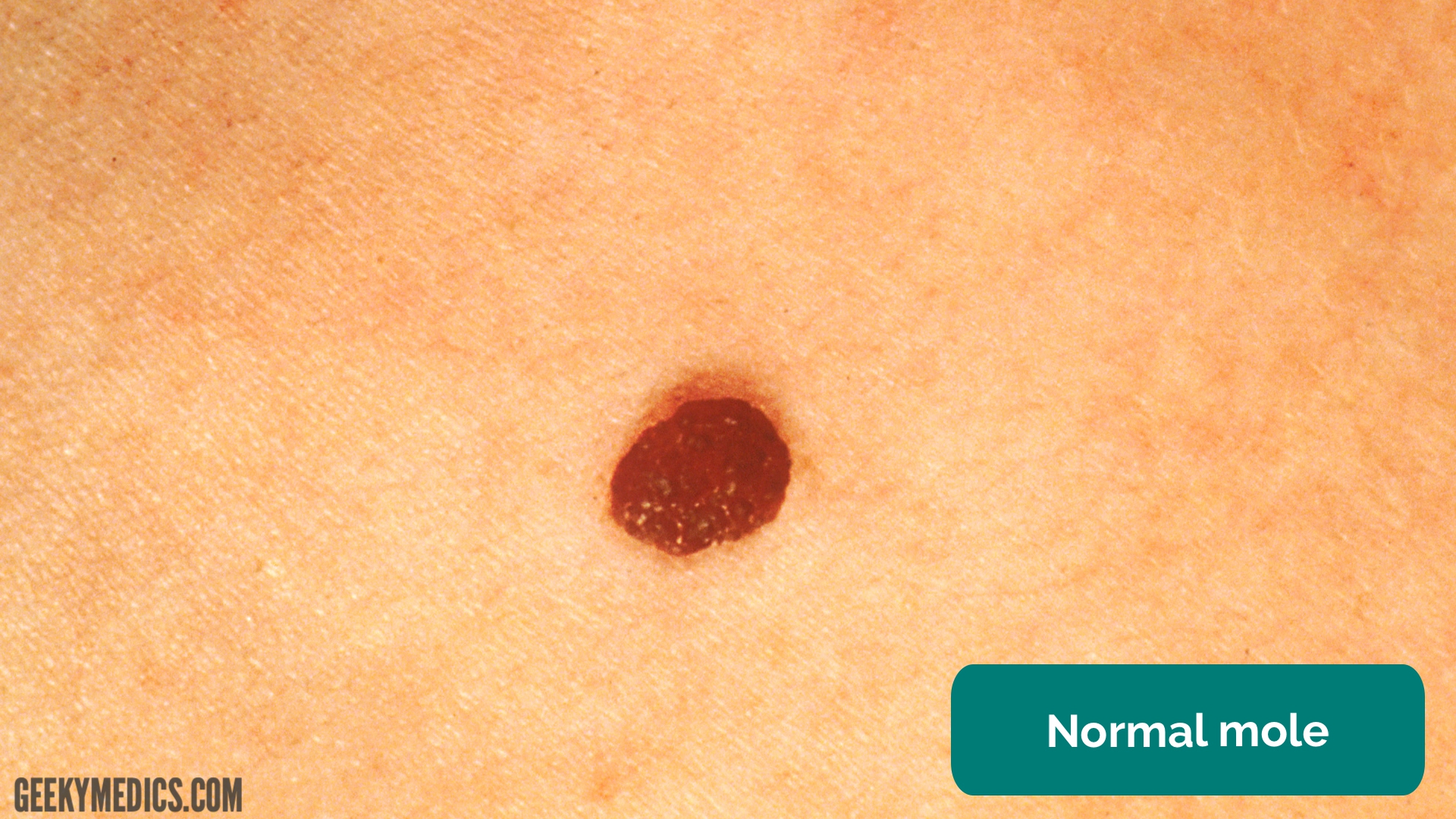
Examining A Skin Lesion Osce Guide Geeky Medics
Flat vascular malformations tend to persist but raised vascular lesions known as hemangiomas generally involute.

Blanching lesion meaning. Non-blanching rashes are caused by small bleeds in the vessels beneath the skin giving a purplish discolouration. It is a characteristic of both purpuric and petechial rashes. They occur due to bleeding beneath the surface of the skin.
Pale and whitish appearance of the skin resulting in uneven skin tone. A non-blanching rash NBR is a skin rash that does not fade when pressed with and viewed through a glass. If the rash disappears or turns white its a blanching rash.
Rashes that blanch when touched arent usually serious. Skin is not broken but is red or discolored or may show changesin hardness or temperature compared to surrounding areas. Blanching of the Skin - Healthline.
Usually skin cancers dont blanch. Feeling of coolness of the skin in the area where the blanching has occurred. The skin changes color slowly over time and is caused by gentle changes in pressure.
Skin lesions that blanch mean the color goes out of them when you press a glass slide on the lesion. You can see that both the blanching and the non-blanching rash look exactly the same without the glass. Cherry angiomas are extremely common benign red blue purple or almost black lesions occurring in middle age on the trunk.
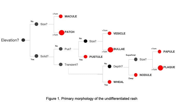
Blanching or not the only way to tell for sure is a biopsy. Paler skin complexion in certain specific parts of the body. If the lesion such as a dark spot on the skin isnt raised and its less than 1 cm in size its by definition a macule.
Most rashes are blanching rashes including virus rashes and allergic reactions. A lesion is any damaged or abnormal area of tissue in the body. Individual purpura measure 310 mm 031 cm 3 32-3 8 in whereas petechiae measure less than 3 mm.
By contrast blanching rashes fade or. A macule can be a variety of colors based on the cause. Blanch medical a temporary whitening of the skin due to transient ischemia Blanching cooking cooking briefly in boiling water Blanching coinage a method used to whiten metal.
Squamous cell carcinomas are usually scaly and basal cell carcinomas look like a nodule with a little bite out of the center. Whitening of the skin when pressure is applied on a certain part of the body. A non-blanching rash can be a symptom of bacterial meningitis but this is not the exclusive cause.
Occasionally they become thrombosed and may fall off or persist as a firm bluish papule. Blanching of skin is defined by the paling or whitening of skin. It forms a dimple when pinched.
Depending on the size of the individual lesions they can be defined as. A skin biopsy shows fibrohistiocytic cell proliferation with entrapment of collagen at the periphery 12. Fading of the skin colour.
Blanching of the skin is typically used by doctors to describe. The way to tell if a rash is blanching or non-blanching is to place a clear drinking glass over the rash and press down. The redness or change in color does not fade within 30.
Although not always necessary. In the French language blanc translates to white Blanching of the skin occurs when the skin becomes white or pale in appearance. Beside above how do I know if I have non blanching rash.
Most lesions are broadly categorized by where they appear in the body for example skin and mouth lesions are some of the most common types. Press glass over rash. Non-blanching rashes are skin lesions that do not fade when a person presses on them.
Dermatofibroma is a reactive lesion that presents as one or more firm dermal papules. This occurs in lesions that get their color from blood supply. If it disappears it is blanching.
They can be easily distinguished from melanocytic lesions by dermoscopy which shows red blue or purple lacunes. 1cm diameter figure 3. The dermatofibroma is pink tan or brown.

Top 5 Causes Of Nonblanching Skin Lesions Clinician S Brief
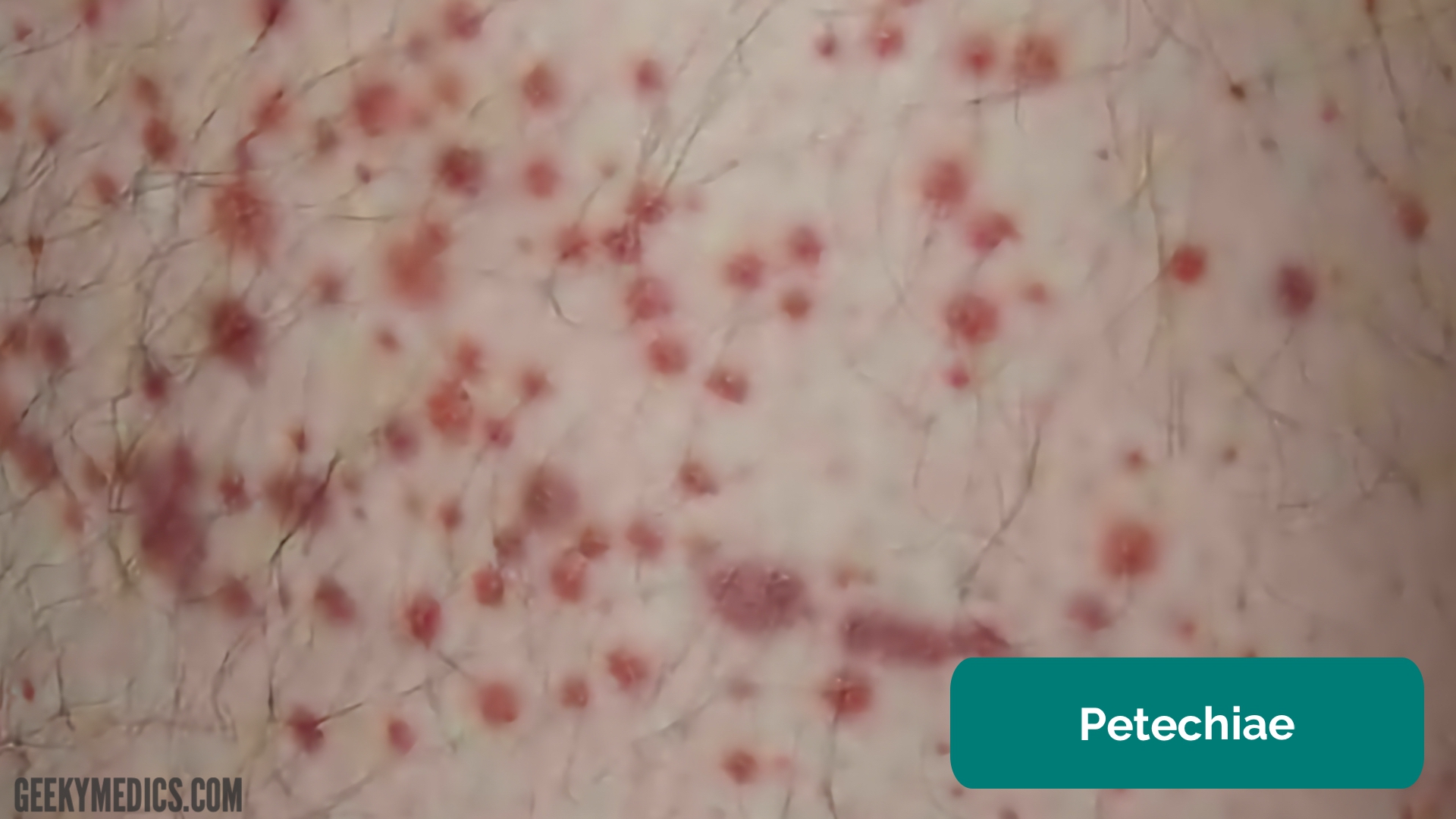
Examining A Skin Lesion Osce Guide Geeky Medics

Top 5 Causes Of Nonblanching Skin Lesions Clinician S Brief

Annular Lesions Diagnosis And Treatment American Family Physician

Pin By Karina Torres On Tattoos Wound Care Nursing Dermatology Nurse Wounds Nursing

Pin By Khubaib Samdani On Medicine Miscellaneous Medical Mnemonics Water Warts Nursing School Tips

Erythematous Oral Lesions When To Treat When To Leave Alone Consultant360
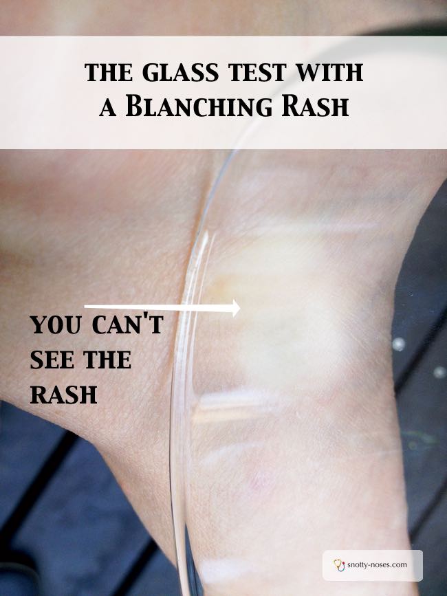
Blanching And Non Blanching Rashes Snotty Noses
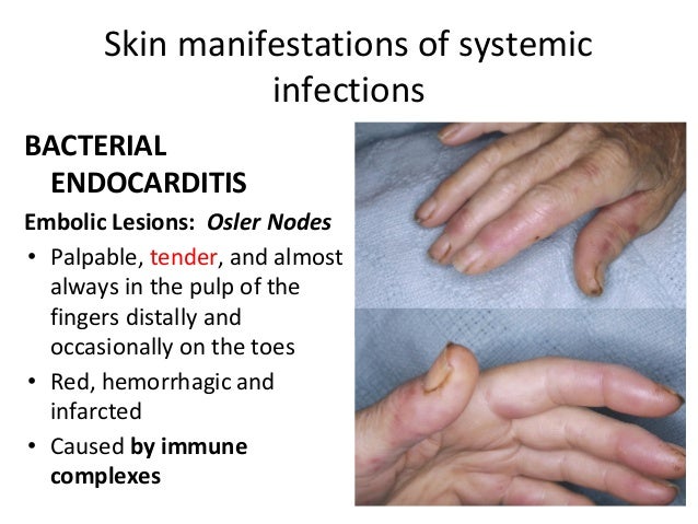
Skin Systemic Infections Final
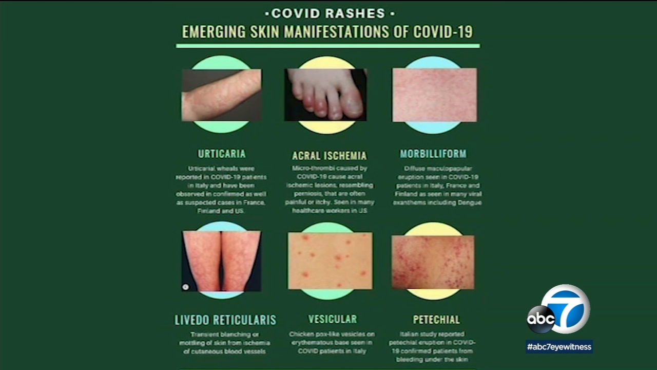
Coronavirus Symptoms Dermatology Organization Issues Guidance On Skin Rashes Associated With Coronavirus Abc7 San Francisco
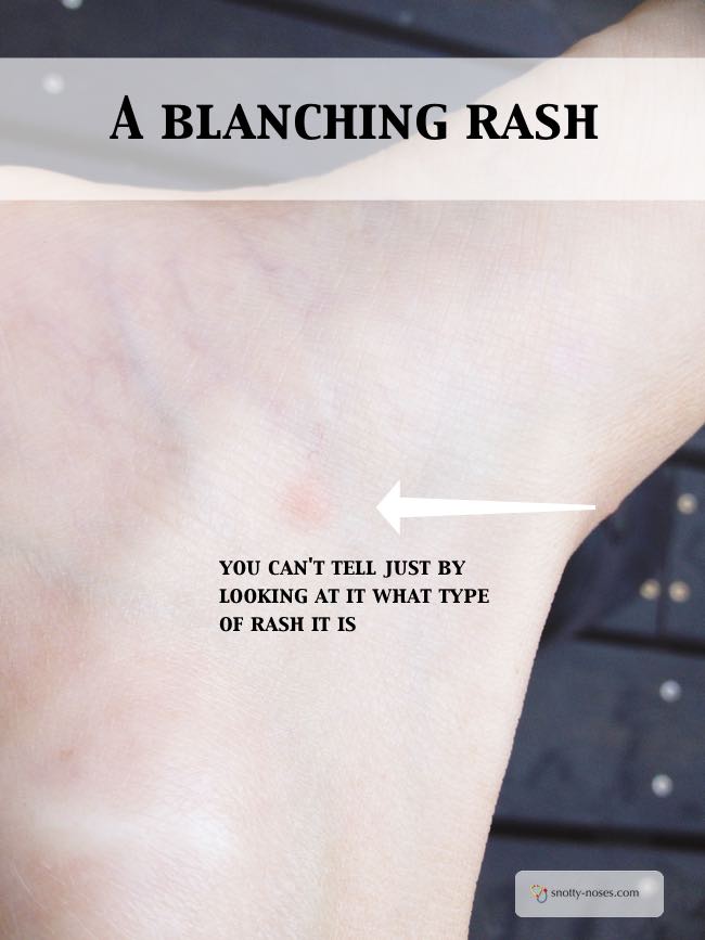
Blanching And Non Blanching Rashes Snotty Noses

Annular Lesions Diagnosis And Treatment American Family Physician

Pemphigus Vulgaris Dermatology Nurse School Nutrition Oral Pathology

Erythema Multiforme Pediatric Nurse Practitioner Pediatric Nursing Dermatology Nurse


Post a Comment for "Blanching Lesion Meaning"